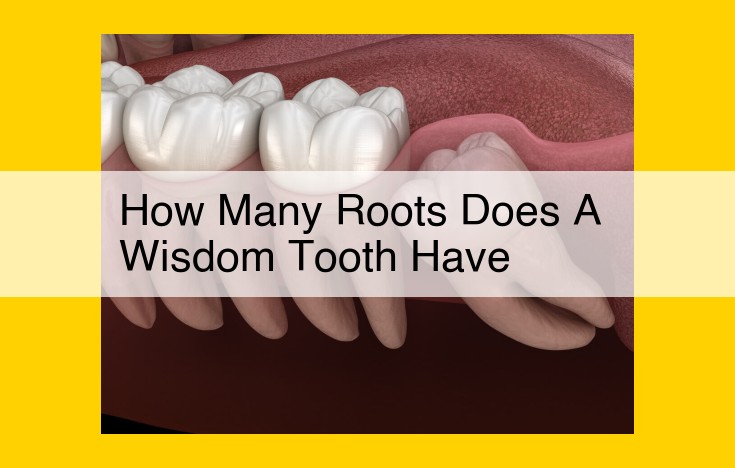
Wisdom teeth, also known as third molars, typically have multiple roots, usually between 2 and 4. These roots can vary in shape and size, with the most common combination being two mesial (towards the front of the mouth) roots and one distal (towards the back of the mouth) root. Understanding the root anatomy of wisdom teeth is crucial for successful dental procedures involving these teeth, such as extractions or root canal treatments.
Delving into the Fascinating World of Tooth Anatomy
Teeth, those pearly whites that adorn our smiles and enable us to savor the culinary delights of life, are marvels of biological engineering. To understand the intricacies of dental procedures and maintain optimal oral health, it’s essential to delve into the realm of tooth anatomy.
Unveiling the Components of a Tooth
Each tooth is a complex structure composed of several distinct components. The crown is the visible portion above the gum line, while the root is the hidden portion anchored within the jawbone. Connecting the crown and root is the neck. Within the tooth lies the pulp cavity, a chamber housing delicate nerves and blood vessels responsible for tooth sensation and nourishment.
Types of Roots: Navigating the Tooth’s Anchorage
Teeth exhibit varied root configurations, each serving a specific purpose. Mesial roots face the front of the mouth, while distal roots face the back. Lingual roots are positioned toward the tongue, and buccal roots are located toward the cheeks. This diverse arrangement provides a firm foundation for teeth to withstand masticatory forces during chewing.
Deciphering Tooth Types: Function and Form
Human dentition comprises four primary tooth types, each tailored to specific functions. Incisors are the chisel-shaped front teeth designed for biting. Canines, with their pointed tips, are responsible for tearing and gripping. Premolars and molars, located in the back of the mouth, serve for grinding and crushing food. Wisdom teeth, often erupting in young adulthood, are the vestigial remnants of our evolutionary past. Understanding the unique characteristics of each tooth type allows us to appreciate the harmonious function of our dental system.
Root Canal Anatomy: Unraveling the Complexities of Dental Structures
Understanding the intricate anatomy of root canals is crucial for successful endodontic treatment, often known as root canal therapy. It involves manipulating and shaping the root canal, which houses the dental pulp, consisting of blood vessels, nerves, and connective tissue.
The shape of root canals varies significantly among teeth. The most common configuration is triradiate, which resembles a triangle with three arms extending from the root’s tip. In some cases, a quadriradiate shape is observed, with four arms instead of three. Understanding these variations is imperative as they influence the difficulty and complexity of endodontic treatment.
The Significance of Root Canal Anatomy in Endodontic Treatment
Grasping the intricacies of root canal anatomy is paramount for endodontic procedures to be effective. During root canal therapy, the inflamed or infected dental pulp must be removed, and the root canal cleaned, shaped, and sealed.
To achieve these objectives, dentists employ specialized instruments to navigate the narrow, often curved, root canals. The shape and number of roots influence the selection of appropriate instruments and techniques, making a thorough understanding of root canal anatomy crucial for optimal treatment outcomes.
Step-by-Step Endodontic Treatment
Endodontic treatment involves a series of meticulous steps:
-
Accessing the Root Canal: The dentist creates an opening through the tooth’s crown to reach the root canals.
-
Cleaning and Shaping the Root Canal: Using specialized files, the dentist removes the infected pulp and enlarges the root canal, preparing it for filling.
-
** obturation:** The cleaned and shaped root canal is filled with a biocompatible material, such as gutta-percha, to seal it and prevent further infection.
Root canal anatomy plays a pivotal role in ensuring the effectiveness of endodontic treatment. Understanding the variations in root canal shapes and their significance are essential for successful outcomes. Through a thorough examination and skilled execution of endodontic procedures, dentists can restore the health and functionality of damaged or infected teeth, providing patients with lasting relief from dental pain.
Imaging Techniques in Dental Anatomy: A Peek into the Intricate World of Teeth
The field of dental anatomy is an intriguing blend of science and artistry, with a profound understanding of tooth structure being paramount for maintaining optimal oral health. To unravel the mysteries that lie beneath the enamel, dental professionals rely on a suite of advanced imaging techniques that provide invaluable insights into tooth and root canal anatomy.
Periapical Radiographs: Capturing the Essence of a Single Tooth
Periapical radiographs, often referred to as “dental X-rays,” are a cornerstone of dental diagnostics. These two-dimensional images offer a precise snapshot of a single tooth, revealing its crown, root, and surrounding bone structure. By exposing the patient to a controlled dose of radiation, periapical radiographs illuminate caries (cavities), cracks, infections, and other abnormalities, enabling dentists to make informed treatment decisions.
Panoramic Radiographs: A Panoramic Vista of the Dental Landscape
Panoramic radiographs take a step back to provide a panoramic view of the entire dentition, including teeth, jaws, and associated structures. This comprehensive image allows dentists to assess the overall health of the mouth, identify impacted or unerupted teeth, detect cysts or tumors, and plan for surgical interventions. Panoramic radiographs are particularly valuable for orthodontic treatment planning, as they reveal the positions and relationships of all teeth within the jaw.
Cone Beam Computed Tomography (CBCT): A 3D Revolution
Cone beam computed tomography (CBCT) represents the cutting-edge of dental imaging technology. This three-dimensional imaging technique utilizes a cone-shaped X-ray beam to produce high-resolution images of the teeth, jaws, and surrounding tissues. CBCT scans provide unmatched detail and depth, enabling dentists to diagnose complex anatomical variations, assess bone density, plan for dental implants, and guide surgical procedures with greater precision.
The Significance of Imaging in Dental Diagnostics and Treatment
Dental imaging techniques play a vital role in diagnosing and treating dental problems. By providing detailed visualizations of tooth and root canal anatomy, these tools empower dentists to:
- Detect hidden decay or infections that may not be visible to the naked eye
- Determine the extent and severity of dental issues
- Plan optimal treatment strategies based on accurate anatomical information
- Monitor the progress of treatment and adjust the plan as needed
- Enhance patient education by visually demonstrating dental anatomy and the effects of various treatments
In conclusion, dental imaging techniques are indispensable for unraveling the intricate world of teeth. Periapical radiographs, panoramic radiographs, and cone beam computed tomography provide a range of diagnostic capabilities, enabling dentists to effectively identify, assess, and treat dental problems, ensuring a healthy and beautiful smile.
Common Dental Treatment Procedures: When Does Your Smile Need Expert Care?
Dental problems are not always minor inconveniences. Sometimes, a simple toothache can signal an underlying issue that requires professional attention. In these cases, common dental treatment procedures like tooth extraction (exodontia) or endodontic treatment (root canal therapy) become necessary to restore your oral health.
Tooth Extraction (Exodontia)
Tooth extraction is a procedure performed to remove a damaged or infected tooth that cannot be saved. It is often the last resort when other treatment options have failed. Indications for tooth extraction include:
- Severe tooth decay or infection that has spread to the pulp or root
- Tooth fracture that cannot be repaired
- Crowding that interferes with the normal alignment of teeth
- Impacted wisdom teeth that are causing pain or discomfort
Endodontic Treatment (Root Canal Therapy)
Root canal therapy is a procedure that involves removing the infected or inflamed pulp from the inside of the tooth. The pulp is the soft tissue that contains nerves, blood vessels, and connective tissue. Root canal therapy is necessary when the pulp becomes infected or inflamed due to:
- Deep tooth decay
- Trauma or injury to the tooth
- Repeated dental procedures on the same tooth
Importance of Proper Diagnosis and Treatment Planning
Both tooth extraction and root canal therapy are serious procedures that should only be performed after a thorough diagnosis and treatment plan. A skilled dentist will consider the following factors:
- The extent and severity of the tooth damage or infection
- The overall health of the patient and their medical history
- The potential risks and benefits of each treatment option
By carefully considering these factors, the dentist can develop a tailored treatment plan that will provide the best possible outcome for the patient’s oral health and overall well-being. If you are experiencing dental problems, don’t hesitate to schedule an appointment with your dentist for an accurate diagnosis and the appropriate treatment plan to restore your smile.
Dental Research and Education: Advancing the Frontiers of Dental Care
The world of dentistry is constantly evolving, thanks to the tireless efforts of researchers and educators dedicated to expanding our knowledge of dental anatomy and morphology. At the forefront of this pursuit are numerous esteemed research centers and organizations, leading the charge in groundbreaking discoveries that shape the future of dental care.
Major Dental Research Centers and Organizations:
- National Institute of Dental and Craniofacial Research (NIDCR): The flagship research institute of the National Institutes of Health, NIDCR is renowned for its groundbreaking research on oral health and diseases, including dental anatomy and root canal morphology.
- American Dental Association (ADA): As the world’s largest dental association, the ADA plays a crucial role in advancing dental research and education. Through its numerous programs and initiatives, it fosters collaboration among researchers and practitioners.
- International Association for Dental Research (IADR): IADR is a global organization dedicated exclusively to dental research. Its annual meetings and publications provide a platform for researchers worldwide to share their latest findings and spur innovation.
Key Textbooks on Dental Anatomy and Histology:
- Ten Cate’s Oral Histology: Development, Structure, and Function by Antonio Nanci and Lester Scott Burstone
- Wheater’s Functional Histology: A Text and Colour Atlas by Barbara Young, James S. Lowe, Alan Stevens, and John W. Heath
- Orban’s Oral Histology and Embryology by G.S. Kumar, R.R. Garg, and M.A. Goyal
These comprehensive textbooks serve as indispensable resources for dental students and practitioners, providing in-depth knowledge of the structure and function of oral tissues.
Online Dental Databases and Journals:
The digital age has revolutionized access to dental research. Online databases such as PubMed and Scopus provide a vast repository of scientific articles and abstracts. Additionally, reputable dental journals like the Journal of Dental Research and Quintessence International disseminate the latest research findings, keeping practitioners abreast of advancements in the field.
By embracing these research and educational resources, dental professionals can stay at the forefront of dental knowledge and provide the highest quality of care to their patients. As the field continues to advance, we can expect even greater breakthroughs that will further improve oral health outcomes.
