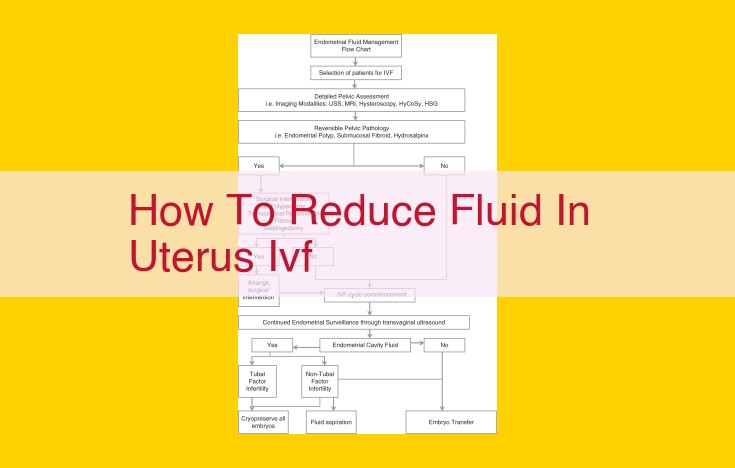
Fluid accumulation in the uterus, known as effusion, can be reduced through various techniques. One is peritoneoscopy, a minimally invasive procedure that allows the surgeon to view the uterus and surrounding organs and remove excess fluid. Another is drainage, where a catheter is inserted to suction out the fluid. Fluid aspiration, using a needle, and paracentesis, which involves inserting a needle through the abdomen, are also used. Understanding the terms “effusion,” “ascites,” and anatomical structures like the uterus and ovaries is crucial for managing these conditions effectively.
Medical Terms:
- Explains the definitions of “effusion” and “ascites,” including their medical significance and common causes.
Medical Terms: Effusion and Ascites
What are effusions and ascites?
In the realm of medicine, effusions refer to abnormal accumulations of fluids within various body cavities. These fluids can be composed of a mix of water, proteins, blood cells, and other substances. Ascites, a specific type of effusion, involves the buildup of fluid in the peritoneal cavity, the space that surrounds your abdominal organs.
Medical Significance
Effusions and ascites are not diseases in themselves but rather symptoms indicative of underlying medical conditions. They can result from a plethora of causes, ranging from infections and inflammation to liver or kidney disorders and even malignancies. Accurate diagnosis is crucial to address the root cause and prevent further complications.
Common Causes
- Abdominal infections like peritonitis can trigger an inflammatory response, leading to the release of fluids into the peritoneal cavity.
- Liver diseases such as cirrhosis or hepatitis can obstruct the normal flow of fluids in the liver, causing ascites.
- Kidney failure impairs the elimination of excess fluids and electrolytes, contributing to effusions and ascites.
- Heart failure can result in fluid retention throughout the body, including within the abdomen (ascites).
- Malignancies can cause effusions or ascites due to tumor growth or obstruction of lymphatic drainage.
Medical Procedures for Managing Effusions and Ascites
When dealing with fluid accumulation in the body, such as pleural effusion or ascites, medical professionals have various procedures at their disposal to manage the condition effectively. These techniques aim to remove excess fluid and prevent complications.
Peritoneoscopy
Peritoneoscopy is a minimally invasive surgical procedure that involves making small incisions in the abdomen to insert a laparoscope, a thin, flexible tube equipped with a camera. The camera provides a clear view of the peritoneal cavity, allowing surgeons to examine the organs and identify the source of the fluid accumulation. Additionally, peritoneoscopy enables the removal of fluid samples for further analysis.
Drainage
In cases of excessive fluid accumulation, drainage may be necessary to relieve pressure and discomfort. Drainage involves inserting a small tube, called a drain, into the cavity where the fluid has accumulated. The drain creates a pathway for the fluid to flow out of the body, reducing the pressure and promoting healing.
Fluid Aspiration
Fluid aspiration is a technique used to remove fluid from a specific body cavity using a needle and syringe. This procedure is typically performed under local anesthesia and is less invasive than peritoneoscopy or drainage. Fluid aspiration provides a quick and efficient way to relieve pressure and obtain fluid samples for diagnostic purposes.
Paracentesis
Paracentesis is a specific type of fluid aspiration used to remove fluid from the peritoneal cavity. This procedure is commonly employed in managing ascites, an accumulation of fluid in the abdomen. Paracentesis involves inserting a needle into the peritoneal cavity and withdrawing the fluid. It can be performed as a diagnostic or therapeutic measure, depending on the underlying cause of the fluid accumulation.
By understanding the purpose and techniques of these medical procedures, individuals with effusions or ascites can be more informed about their treatment options. These procedures play a critical role in managing fluid accumulation, relieving symptoms, and preventing complications, ultimately improving the quality of life for patients.
Anatomical Structures Associated with Effusions and Ascites
When discussing effusions and ascites, it’s crucial to understand the anatomical structures involved and their role in these conditions.
Uterus: The uterus is the primary female reproductive organ, responsible for carrying a developing fetus. Ascites can accumulate in the pelvis and uterus, often due to blockages or infections that prevent normal fluid drainage.
Ovaries: These small, bean-shaped organs attach to the uterus and produce eggs. Ovarian cysts or tumors can interfere with fluid drainage, leading to effusions and ascites.
Cul-de-sac: This is the deepest part of the pelvic cavity, located behind the uterus and in front of the rectum. Effusions can accumulate in the cul-de-sac, creating a fluid-filled pocket known as a pouch of Douglas.
Cervix: The cervix is the lower part of the uterus that connects to the vagina. Cervical cancer or other abnormalities can obstruct fluid flow, contributing to effusions and ascites.
Understanding these anatomical structures and their role in effusions and ascites helps guide proper diagnosis and management of these conditions.
