3. The Thoracic Cage and the Axial Skeleton
- Define the axial skeleton and its components
- Explain the connection between the thoracic cage and other parts of the axial skeleton
- Discuss the role of the thoracic cage in protecting the vital organs
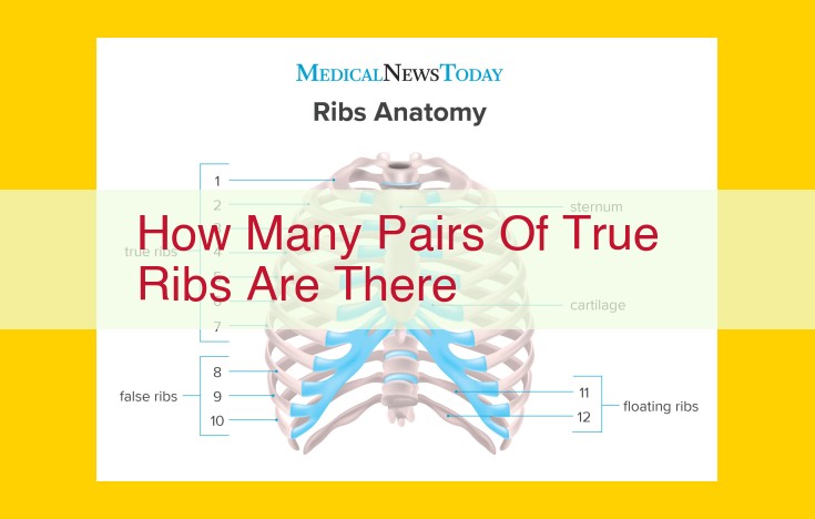
- **How many pairs of true ribs are there?**
Unveiling the Thoracic Cage: A Journey into the Human Chest
Embark on a storytelling adventure to explore the enigmatic thoracic cage, the intricate framework that encases our vital organs.
Deep within the human torso resides a remarkable structure known as the thoracic cage, a complex yet elegant assembly of bones and cartilages. Nestled between the flexible neck above and the robust abdomen below, it forms the foundation of our respiratory and circulatory systems.
The Thoracic Cage: A Haven for Vital Organs
Imagine a protective vault, meticulously crafted to safeguard the most precious treasures. The thoracic cage serves this very purpose, shielding our delicate heart, lungs, and major blood vessels from harm. Its sturdy walls, composed of the sternum, costal cartilages, and intercostal muscles, work in unison to provide unwavering support and protection.
A Vital Player in Respiration: The Thoracic Cage’s Dance of Life
With every breath we take, the thoracic cage orchestrates a rhythmic dance that sustains our very existence. During inhalation, the intercostal muscles contract, drawing the ribcage outward and upward, while the diaphragm descends, further enlarging the chest cavity. This expansion creates a vacuum that draws air into the lungs.
As we exhale, the thoracic cage reverses its motion. The intercostal muscles relax, allowing the ribcage to return to its resting position, and the diaphragm rises, pushing air out of the lungs. This intricate interplay ensures the constant exchange of oxygen and carbon dioxide that fuels our bodies.
The Thoracic Cage’s Axial Connection: A Symphony of Bones
The thoracic cage forms an integral part of the axial skeleton, the central support system of our body. It articulates with the spine, ribs, and sternum, creating a stable and interconnected framework. This seamless integration provides structural stability, enabling us to stand, walk, and perform countless other movements.
Moreover, the thoracic cage plays a pivotal role in protecting the spinal cord, a crucial nerve bundle that transmits messages between the brain and the rest of the body. This protective enclosure ensures the uninterrupted flow of information that governs our every action and thought.
The thoracic cage, with its intricate design and vital functions, stands as a testament to the marvels of human anatomy. It is a symphony of bones, cartilages, and muscles that work together to safeguard our essential organs, enable respiration, and provide structural support. As we delve deeper into its secrets, we gain a profound appreciation for its indispensable role in the human experience.
Three main components: sternum, costal cartilages, and intercostal muscles
The Thoracic Cage: A Symphony of Bones and Cartilage
Nestled amidst our rib cage lies the thoracic cage, a remarkable structure composed of three main components that form a protective shield around our vital organs.
The sternum, a flat bone, forms the front of the thoracic cage. It articulates with the ribs, providing stability and protecting the heart and lungs. Connecting the ribs to the sternum are the costal cartilages, flexible bands of tissue that allow for chest expansion during breathing.
Lastly, the intercostal muscles, located between the ribs, play a crucial role in respiration. By contracting and relaxing, these muscles lift and lower the ribs, expanding and contracting the thoracic cage to facilitate the flow of air.
The Thoracic Cage: A Vital Part of Respiration
The thoracic cage is an indispensable component of the respiratory system. When we inhale, our diaphragm contracts, pulling the thoracic cavity downward. This motion causes the intercostal muscles to contract, lifting the ribs upward and expanding the chest cavity. As the chest cavity expands, air is drawn into the lungs.
Upon exhalation, the intercostal muscles relax, lowering the ribs and reducing the volume of the chest cavity. This forces air out of the lungs through the breathing passages.
The Thoracic Cage: Protecting the Vital Organs
The thoracic cage forms part of the axial skeleton, the central framework of our body. It connects with the spine, shoulder blades, and collarbones, providing support and stability. As a protective barrier, the thoracic cage safeguards the heart, lungs, and other vital organs from external forces and injuries.
By understanding the anatomy and functions of the thoracic cage, we appreciate the intricate design of our bodies and the vital role it plays in our survival. From its role in respiration to its protective function, the thoracic cage is a testament to the marvel of human physiology.
The Thoracic Cage’s Vital Role in Respiration
The thoracic cage, the protective structure that houses our vital organs, is not merely a passive vessel. It plays a dynamic role in the intricate process of respiration, guiding the harmonious dance of oxygen intake and carbon dioxide expulsion.
Imagine a symphony orchestra where the thoracic cage serves as the conductor. The muscles between the ribs act as the nimble bows, orchestrating the expansion and contraction of the chest cavity. As the diaphragm, our muscular floor, descends, it draws the rib cage upward, enlarging the cavity. Conversely, when the diaphragm relaxes, the rib cage recoils, reducing the cavity’s volume.
This rhythmic expansion and contraction create a pressure gradient, a force that drives the flow of air in and out of the lungs. When the chest cavity expands, the pressure inside decreases, drawing air into the lungs like a gentle breeze. As the cavity contracts, the pressure rises, expelling carbon dioxide-laden air from the lungs.
Through this intricate interplay, the thoracic cage becomes an integral part of our respiratory system, ensuring our every breath. It is a testament to the body’s remarkable design, where seemingly passive structures play vital roles in sustaining life.
Describe how the chest cavity is enlarged and contracted during breathing
The Thoracic Cage: A Vital Structure for Life’s Symphony
Every breath we take is a testament to the incredible complexity and harmony of our bodies. At the core of this symphony of respiration lies the thoracic cage, a remarkable structure that plays a pivotal role in inhaling and exhaling.
Imagine the thoracic cage as a protective enclosure for your vital organs. Composed of the sternum, ribs, and intercostal muscles, this framework surrounds the heart, lungs, and major blood vessels. When you breathe, this cage expands and contracts, creating the negative pressure necessary for air to rush into your lungs.
As the diaphragm, a muscle separating the thoracic cavity and abdomen, contracts, it pulls the lungs downward. Simultaneously, the intercostal muscles between the ribs contract, lifting and pulling the rib cage outward. This coordinated action enlarges the chest cavity, creating a lower pressure within.
With the chest cavity now expanded, air rushes into the lungs, propelled by the pressure difference between the atmosphere and the lower pressure within. The newly inhaled air carries life-sustaining oxygen to every cell in your body.
As you exhale, the process reverses. The diaphragm relaxes, reducing the volume of the chest cavity. The intercostal muscles relax as well, allowing the rib cage to descend. This contracts the chest cavity, increasing the pressure within and forcing air out of the lungs.
This rhythmic expansion and contraction of the thoracic cage is a symphony of life, ensuring that your body receives the oxygen it needs and expels carbon dioxide. Without this silent guardian, we would cease to breathe, and life’s symphony would end.
The Thoracic Cage: A Protective Haven for Vital Organs
Hidden beneath our ribs lies an essential structure that plays a crucial role in our survival – the thoracic cage. Let’s embark on a journey to unravel the mysteries of this intricate framework.
Chapter 1: The Thoracic Cage – An Architectural Masterpiece
The thoracic cage is a rigid enclosure formed by the sternum, costal cartilages, and intercostal muscles. It envelops the heart, lungs, and other vital organs within the chest cavity, providing them with both support and protection.
Chapter 2: A Dynamic Symphony – The Thoracic Cage and Respiration
The thoracic cage is an active participant in our breathing process. When we inhale, the intercostal muscles contract, expanding the chest cavity and lowering the diaphragm. This creates a negative pressure that draws air into the lungs. Conversely, exhalation occurs when the intercostal muscles relax, allowing the diaphragm to rise and push air out.
Chapter 3: The Axial Skeleton – A Backbone of Support
The axial skeleton is the core structure that forms our body’s central axis. It comprises the skull, vertebral column, and thoracic cage. These components work together to provide stability, support, and protection for the spinal cord, brain, and other vital organs.
Chapter 4: The Thoracic Cage – A Vital Link
The thoracic cage plays a pivotal role in linking the axial skeleton to other skeletal elements. It connects to the cervical vertebrae in the neck and the lumbar vertebrae in the lower back, ensuring proper alignment and mobility. This complex network allows for essential movements like bending, twisting, and reaching.
The thoracic cage is more than just a skeletal structure – it’s a vital fortress guarding our most precious organs. Its involvement in respiration and its connection to the axial skeleton underscore its fundamental role in our health and well-being. So, let’s appreciate this anatomical marvel that tirelessly works behind the scenes to keep us breathing and moving with ease.
The Thoracic Cage: A Vital Component of Our Skeletal System
The Thoracic Cage: A Protective Barrier
Nestled within the upper portion of our body, the thoracic cage is an intricate structure composed of the sternum, costal cartilages, and intercostal muscles, forming a protective shield around our vital organs. The ribs, connected to the vertebrae at the back and the sternum at the front, create a rigid framework that safeguards the heart, lungs, and major blood vessels.
The Thoracic Cage: A Mechanism for Breathing
This remarkable structure is not merely a passive enclosure. It plays a pivotal role in our respiratory system. As we inhale, the diaphragm contracts and the intercostal muscles elevate the ribs, expanding the chest cavity. This creates a negative pressure that draws air into the lungs. Conversely, exhalation occurs when the diaphragm relaxes and the intercostal muscles lower the ribs, reducing the volume of the chest cavity and expelling air.
The Thoracic Cage: A Part of the Axial Skeleton
The thoracic cage forms an integral part of the axial skeleton, which includes the skull, vertebral column, and rib cage. The vertebrae, stacked one upon another, provide structural support and house the spinal cord. The ribs, extending from the vertebrae, connect to the sternum anteriorly, forming the thoracic cage. Together, these components provide stability and protect our spinal cord and internal organs.
The Thoracic Cage: A Sentinel of Our Health
The thoracic cage is not only an anatomical marvel but also a sentinel of our health. Abnormalities in its shape or function can indicate underlying medical conditions. A deformed thoracic cage, for example, may suggest congenital defects or respiratory problems. Similarly, asymmetrical rib movement can be a sign of lung disease or neuromuscular disorders. By understanding the structure and function of the thoracic cage, we can better appreciate its role in safeguarding our well-being and maintaining our respiratory health.
Discuss the role of the thoracic cage in protecting the vital organs
The Thoracic Cage: A Shield for Your Vital Organs
Your thoracic cage is a remarkable structure that houses and safeguards your vital organs. Comprised of the sternum, costal cartilages, and intercostal muscles, this protective cage plays a crucial role in maintaining your well-being.
As you inhale, your thoracic cage expands, drawing air into your lungs. This expansion is facilitated by the contraction of your intercostal muscles, which lift the ribs and pull the sternum forward. As you exhale, the thoracic cage contracts, pushing air out of your lungs. This intricate mechanism ensures that your body receives the oxygen it needs while expelling carbon dioxide.
Beyond its respiratory function, the thoracic cage also serves as a protective shield for your delicate organs. The ribs form a strong, bony framework that encases the heart, lungs, and other vital structures. The sternum, a flat bone located at the front of the chest, provides additional support and protection.
The vertebral column, which forms the backbone, connects to the thoracic cage at the rib joints. This connection stabilizes the cage and allows for movement of the trunk. The thoracic cage, therefore, plays a vital role in both respiration and protection, ensuring the proper functioning and safety of your essential organs.
