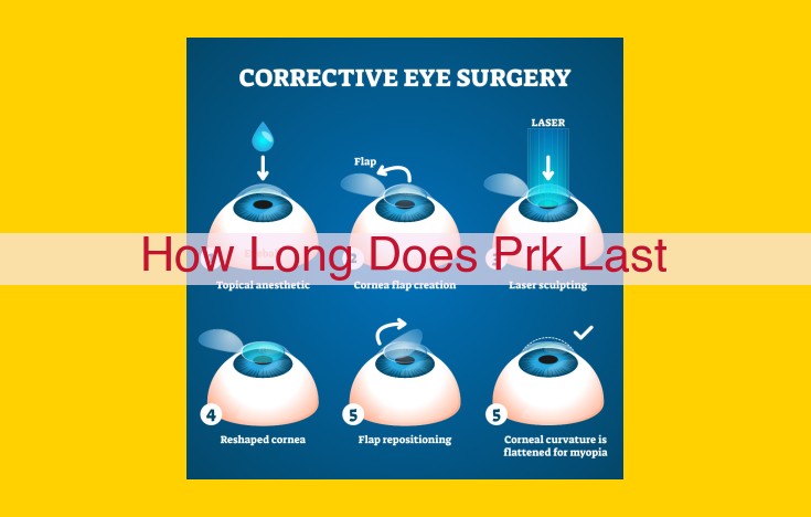
While PRK can provide permanent vision correction, the longevity of its effects can vary depending on individual factors. Generally, most patients experience stable vision for several years to decades. However, factors such as corneal healing, lifestyle, and underlying eye conditions can influence the duration of the results. Regular eye exams are essential to monitor vision stability and address any potential changes.
Refractive Surgery: Photorefractive Keratectomy (PRK)
Understanding the Key Structures of the Eye Involved in PRK
Photorefractive Keratectomy (PRK) is a refractive surgery procedure that aims to improve vision by reshaping the cornea, the transparent outer layer of the eye. To fully grasp the essence of this procedure, it’s essential to understand the critical structures within the eye involved in PRK.
At the forefront lies the corneal epithelium, a thin layer of non-living cells covering the cornea. Beneath it resides the stromal layer, the cornea’s thickest layer, composed of collagen fibers that give the cornea its shape. PRK primarily targets the corneal stroma to reshape it and correct vision abnormalities.
Positioned beneath the stroma is Bowman’s layer, a thin, transparent membrane that separates the stroma from the Descemet’s membrane, another delicate layer located near the back of the cornea. The innermost layer of the cornea is the corneal endothelium, a single layer of cells responsible for maintaining corneal hydration.
How the Cornea’s Anatomy Impacts PRK
Photorefractive keratectomy (PRK) involves reshaping the cornea, the transparent outer layer of the eye, to correct refractive errors. To understand PRK’s mechanism, let’s scrutinize the cornea’s intricate anatomy and its harmonious interplay during the procedure.
Epithelium: The Outer Guard
The outermost layer of the cornea, the epithelium, acts as a protective barrier against external threats. During PRK, it’s meticulously removed to unveil the underlying stroma.
Stroma: The Structural Heart
The stroma, the cornea’s thick middle layer, comprises intricate collagen fibers that lend it its exceptional strength. Excimer lasers, during PRK, precisely ablate the stroma, reshaping its curvature to correct refractive defects.
Bowman’s Layer: The Resilient Defender
Nestled beneath the epithelium, Bowman’s layer, a thin but hardy membrane, shields the stroma from external forces. It provides stability as the laser meticulously resculpts the cornea.
Descemet’s Membrane: The Inner Barrier
Descemet’s membrane, a delicate layer on the cornea’s inner surface, plays a crucial role in maintaining the eye’s intraocular pressure. During PRK, it remains untouched, ensuring the cornea’s structural integrity.
Endothelium: The Vital Lining
The endothelium, the innermost layer of the cornea, is responsible for pumping excess fluid out of the cornea. Preserving its integrity is essential for maintaining the cornea’s clarity and overall health.
Describe the excimer laser, its function in PRK, and its advantages over other lasers.
The Excimer Laser: Unveiling the Precision of PRK**
In the realm of refractive surgery, the excimer laser stands as a beacon of precision, reshaping the corneal landscape with unmatched accuracy. This ultraviolet laser holds the key to the success of photorefractive keratectomy (PRK), a procedure that corrects vision by sculpting the cornea.
Unlike other lasers, the excimer laser emits light at a specific wavelength that is absorbed by the corneal stroma, the thickest layer of the cornea. This absorption creates a precise ablation, removing a microscopic amount of tissue with each pulse. The result is a reshaped corneal surface that directs light more accurately onto the retina, restoring clear vision.
The advantages of the excimer laser over other lasers are undeniable. Its precise ablation allows for customized treatments tailored to each patient’s unique eye conditions. Additionally, the laser’s minimal thermal effect preserves the corneal tissue surrounding the treatment area, maintaining its integrity and minimizing complications.
Moreover, the excimer laser’s ability to create smooth corneal surfaces reduces the risk of optical aberrations and halos around lights at night. This results in excellent visual outcomes with high patient satisfaction.
So, as you embark on your journey toward clear vision with PRK, trust in the precision of the excimer laser, the technological marvel that will restore your sight to its full potential.
Wavefront Technology and the Customization of PRK Treatments
In the realm of refractive surgery, Photorefractive Keratectomy (PRK) stands tall as an exceptional procedure for correcting refractive errors. As with any surgical technique, precision plays a paramount role in ensuring optimal outcomes. This is where wavefront technology steps into the spotlight.
Wavefront technology** is an advanced optical measurement system that maps the unique imperfections of an individual’s cornea. It does this by sending a beam of light through the eye and measuring how it distorts. This detailed mapping allows surgeons to create a customized treatment plan for each patient, taking into account their unique corneal shape and refractive errors.
By utilizing wavefront-guided PRK, surgeons can precisely reshape the cornea to correct refractive errors, including nearsightedness, farsightedness, and astigmatism. The procedure involves using an excimer laser to remove a thin layer of corneal tissue, reshaping it to focus light properly onto the retina. The customized treatment plan ensures that the cornea is reshaped to the ideal curvature, minimizing aberrations and maximizing visual clarity.
The precision offered by wavefront technology results in numerous benefits for PRK patients:
- Reduced risk of complications
- Enhanced visual acuity
- Faster visual recovery times
- Improved night vision
- Reduced dependence on corrective eyewear
As a result, wavefront technology has revolutionized PRK, allowing surgeons to deliver highly customized treatments that restore patients’ vision to its full potential.
Refractive Surgery: Photorefractive Keratectomy (PRK)
Personnel and Considerations
The Role of the Ophthalmologist in PRK
In the realm of PRK, the ophthalmologist holds the reins as the expert surgeon responsible for executing the procedure with precision and care. These highly skilled professionals possess years of specialized training and experience in diagnosing and treating eye conditions, ensuring your vision is in the safest hands.
Before embarking on PRK, a thorough preoperative evaluation is crucial. The ophthalmologist meticulously employs cutting-edge technology to assess the corneal topography (the shape and thickness of your cornea), refraction (the ability of your eyes to focus light), and pupil dilation (enlarging the pupils for a comprehensive examination).
Armed with these insights, the ophthalmologist carefully designs a personalized PRK treatment plan tailored specifically to your unique visual needs. During the procedure, they deftly employ the excimer laser, skillfully reshaping your cornea to correct any refractive errors.
Postoperatively, the ophthalmologist diligently monitors your progress, providing expert guidance and meticulous care. They prescribe pain management medication to alleviate discomfort, administer antibiotics to prevent infection, and provide detailed instructions on how to protect your precious vision during the healing process.
Trusting your eyes to a qualified and experienced ophthalmologist is paramount for a successful PRK outcome. Their expertise and dedication ensure that you embark on a journey towards clearer vision with confidence and peace of mind.
Refractive Surgery: Photorefractive Keratectomy (PRK)
1. Key Entities and Their Relevance
Before delving into the technical aspects of PRK, let’s familiarize ourselves with the key structures that play a pivotal role in this procedure.
The cornea, the transparent outermost layer of the eye, consists of several layers. The corneal epithelium, the outermost layer, is removed during PRK. Beneath the epithelium lies Bowman’s layer, followed by the stroma, the thickest layer. Descemet’s membrane and the corneal endothelium form the innermost layers.
2. Technology and Procedures
The excimer laser lies at the heart of PRK, emitting precise pulses of ultraviolet light that reshape the corneal stroma. This laser offers unmatched precision compared to other lasers, allowing for highly customized treatments.
Wavefront technology takes PRK to a new level. It analyzes the unique imperfections of each individual’s eye, enabling the laser to correct aberrations that may have caused blurry vision. This personalized approach enhances the accuracy and outcomes of PRK.
3. Personnel and Considerations
Ophthalmologists with specialized training and expertise are the key personnel performing PRK. Their meticulous assessments ensure the suitability and safety of the procedure.
Preoperative Evaluation
Before surgery, the ophthalmologist conducts a thorough preoperative evaluation involving the following steps:
- Corneal topography: A 3D map of the cornea is created to assess its shape and curvature. This information helps guide the laser treatment.
- Refraction: This non-invasive test measures the eye’s refractive error, determining the power of glasses or contact lenses needed.
- Pupil dilation: Eye drops are used to widen the pupils, allowing for a clearer view of the cornea during surgery.
Postoperative Care: A Crucial Step for Optimal Results
Pain Management
After PRK, you may experience some discomfort, including stinging, burning, and aching. To alleviate this pain, your ophthalmologist will prescribe pain-relieving eye drops or oral medications. It’s essential to follow their instructions and take the medication as directed.
Antibiotics
To prevent infection, your ophthalmologist will prescribe antibiotic eye drops. These drops will combat bacteria that may have entered the eye during surgery. It’s crucial to use the antibiotic eye drops regularly and for the full duration of the treatment.
Eye Protection
After PRK, your eyes will be sensitive to light. To protect them, wear sunglasses or eye shields consistently when outdoors. Additionally, avoid rubbing or touching your eyes, as this can increase the risk of infection and damage the healing process.


