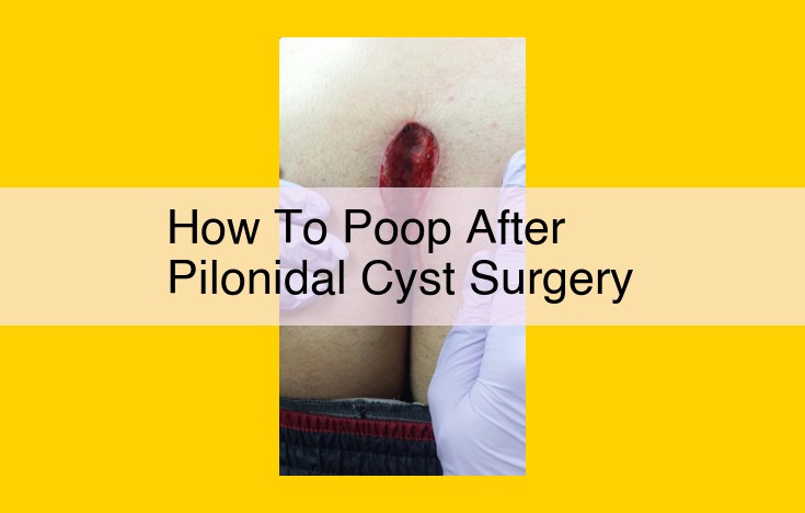
After pilonidal cyst surgery, maintaining bowel regularity is crucial. It is important to follow a high-fiber diet and drink plenty of fluids to avoid constipation. Pain medication may delay bowel movements, so consider taking stool softeners or laxatives as prescribed by your doctor. When using the toilet, utilize a squat position or footstool to elevate your knees, reducing pressure on the surgical site.
Pilonidal Cyst: An Overview of Symptoms, Causes, and Risk Factors
Nestled in the sacrococcygeal region at the base of the spine, a pilonidal cyst is an uncomfortable and potentially painful condition that affects many individuals. Understanding the definition, symptoms, causes, and risk factors of this cyst is crucial for informed decision-making and effective treatment.
Definition
A pilonidal cyst is a small sac filled with hair and debris that forms in the skin near the top of the buttocks. It’s often painful and may drain a foul-smelling fluid.
Symptoms
The most common symptom of a pilonidal cyst is pain. This pain can range from mild to severe and may be worse when sitting or lying down. Other symptoms may include:
– Swelling
– Redness
– Drainage
– Fever
– Chills
Causes
The exact cause of pilonidal cysts is not fully understood, but several factors may contribute to their development:
– Hair growth: Ingrown hairs can irritate the skin and lead to the formation of a cyst.
– Friction: Repeated rubbing against the skin, such as from sitting or tight clothing, can increase the risk of developing a cyst.
– Obesity: Excess weight puts pressure on the sacrococcygeal region, contributing to cyst formation.
Risk Factors
Certain factors increase the likelihood of developing a pilonidal cyst:
– Family history: Having a family member with a pilonidal cyst increases the risk.
– Gender: Men are more likely to develop pilonidal cysts than women.
– Occupation: Jobs that require prolonged sitting or heavy lifting can increase the risk.
– Hygiene: Poor hygiene, such as not showering regularly, can contribute to cyst development.
Surgery for Pilonidal Cyst: A Comprehensive Guide
Understanding the Surgical Options
When conservative treatments fail to alleviate the discomfort and recurrence of a pilonidal cyst, surgery is often the recommended course of action. There are two main surgical procedures used to treat pilonidal cysts:
- Closed excision: This involves excising the cyst, leaving the wound to heal naturally.
- Open excision and reconstruction: This technique entails excising the cyst along with the surrounding sinus tracts. The wound is then left open to heal by secondary intention.
Pre-Operative Preparations
Before surgery, your doctor will conduct a thorough examination to assess the extent of the cyst and determine the appropriate surgical approach. They will also provide detailed instructions on how to prepare for the procedure, which may include:
- Fasting for a certain period before surgery
- Stopping smoking
- Antibiotic prophylaxis
Post-Operative Care
After surgery, your doctor will provide specific guidelines for post-operative care to ensure a successful recovery. These guidelines may include:
- Wound Management: Keeping the surgical site clean and dry is crucial. You will be instructed on how to clean and dress the wound regularly.
- Pain Management: Medications, such as pain relievers and antibiotics, may be prescribed to manage pain and prevent infection.
- Activity Restrictions: Following surgery, you may be advised to avoid strenuous activities and maintain a comfortable sitting position to minimize pressure on the surgical site.
Tips for Smooth Recovery
- Follow your doctor’s instructions carefully to promote optimal healing.
- Contact your doctor immediately if you experience any signs of infection, such as fever, redness, or drainage.
- Be patient and allow ample time for the surgical site to heal completely.
- Consider consulting with a physical therapist for guidance on managing pain and restoring mobility.
Remember, while pilonidal cyst surgery can effectively resolve the discomfort, proper post-operative care is essential for a successful recovery. By following your doctor’s instructions and taking care of the surgical site, you can minimize the risk of complications and promote a swift and comfortable healing process.
Postoperative Care: A Guide to Recovery from Pilonidal Cyst Surgery
Wound Management:
After surgery, managing the surgical site is crucial for proper healing. Keep the wound clean by gently washing it with soap and water twice a day. Regularly change the dressing to prevent infection and promote drainage. Monitor the wound for any signs of infection, such as redness, swelling, or discharge.
Pain Management:
Pain is a common aspect of post-surgical recovery. Your doctor may prescribe pain medication to alleviate discomfort. Non-pharmacological methods, such as ice packs, heating pads, or over-the-counter pain relievers, can also provide relief. Elevate your legs to reduce swelling and promote healing.
Activity Restrictions:
Following surgery, it’s essential to limit activities that could strain the surgical site. Avoid sitting for prolonged periods and engage in light exercise as recommended by your doctor. Listen to your body and rest when necessary to facilitate recovery. Adhering to these guidelines will help minimize pain, prevent complications, and promote optimal healing.
Wound Care: A Crucial Step in Pilonidal Cyst Surgery Recovery
After undergoing surgery for a pilonidal cyst, proper wound care is essential for ensuring a successful and speedy recovery. Here’s a comprehensive guide to help you navigate this important step:
Cleaning the Surgical Site
- Clean the wound gently with warm water and a mild soap or antiseptic solution.
- Use a cotton swab or gauze to gently remove any crusted material around the wound edges.
- Avoid using harsh scrubs or rubbing the wound, as this can irritate and delay healing.
Dressing the Wound
- Apply a fresh dressing over the wound as directed by your doctor.
- Use a sterile gauze pad or dressing to absorb any drainage.
- Change the dressing regularly, typically once or twice a day, to keep the wound clean and prevent infection.
Monitoring for Infection
- Pay attention to the appearance of the wound. It should gradually heal, with minimal redness or swelling.
- Watch for signs of infection, such as:
- Increased redness, swelling, or warmth around the wound
- Discharge that is green, yellow, or foul-smelling
- Fever
- Chills
- Contact your doctor immediately if you experience any signs of infection.
Other Wound Care Tips
- Keep the wound elevated to promote drainage and reduce swelling.
- Avoid wearing tight-fitting clothing that may put pressure on the wound.
- Get plenty of rest to support your body’s natural healing process.
- Follow your doctor’s instructions carefully regarding activity restrictions and medication use.
By adhering to these wound care guidelines, you can minimize the risk of infection and facilitate a smooth recovery from pilonidal cyst surgery.
Pain Management After Pilonidal Cyst Surgery
Understanding the Pain
Following pilonidal cyst surgery, you may experience some level of discomfort and pain. This is a normal part of the healing process as the surgical site recovers. The pain is typically localized to the sacrococcygeal region, where the cyst was removed.
Medications for Relief
Your doctor may prescribe pain relievers such as non-steroidal anti-inflammatory drugs (NSAIDs) or opioid painkillers to manage the discomfort. These medications work by reducing inflammation and blocking pain signals. It’s crucial to follow the prescribed dosage and instructions carefully.
Other Pain Relief Techniques
In addition to medications, there are several non-pharmacological techniques that can provide pain relief. Cold compresses applied to the surgical site can help reduce swelling and numb the pain. Sitz baths with warm water can promote circulation and soothe discomfort. Resting and elevating the affected area can also minimize pain by reducing pressure and strain.
Non-Pharmacological Approaches
For those seeking alternative pain management methods, acupuncture and massage therapy have been shown to effectively reduce pain and improve mobility. Yoga and stretching exercises can also enhance flexibility and alleviate muscle tension, which can contribute to pain.
Importance of Communication
Pain management is an essential aspect of post-operative care. If you experience severe or persistent pain, it’s important to communicate this to your doctor. They can adjust medications, recommend additional pain relief techniques, or investigate any underlying complications. Effective pain management allows you to rest comfortably and support the healing process.
The Sacrococcygeal Region: A Hidden Anatomy and Its Role in Pilonidal Cysts
Nestled deep within the lower back, the sacrococcygeal region is an anatomical landscape often shrouded in mystery. This intriguing area connects the sacrum, a triangular bone at the base of the spine, to the coccyx, a tail-like bone at the very bottom. Its hidden crevices and unique anatomy play a pivotal role in the development of pilonidal cysts, a common condition that affects this region.
Imagine a tiny cleft, a cleft sinus, tucked away within the fold of skin above the coccyx. This cleft is a remnant of our evolutionary past, a reminder of a tail that our ancestors once possessed. In some individuals, this cleft may fail to close completely, creating a small opening that can become infected and inflamed, leading to the formation of a pilonidal cyst.
The sacrococcygeal region is particularly prone to developing pilonidal cysts due to its unique anatomy. The cleft sinus is often located in an area where sweat, hair, and dirt can easily accumulate, creating an ideal environment for bacteria to thrive. Additionally, the constant movement and friction in this region can irritate the cleft sinus, further increasing the risk of infection.
Understanding the anatomy of the sacrococcygeal region and its role in pilonidal cysts is crucial for both prevention and treatment. Proper hygiene, avoiding tight-fitting clothing, and maintaining a healthy weight can help reduce the risk of developing this condition. In cases where a pilonidal cyst does form, prompt medical attention is essential to ensure proper drainage and wound care, preventing further complications and ensuring a comfortable recovery.
The Sacrum: A Key Player in Pilonidal Cyst Development
Nestled at the base of the spine, the sacrum is a triangular bone that forms the posterior wall of the sacrococcygeal region. It plays a vital role in the development of pilonidal cysts, a condition that affects the lower back.
The sacrum’s unique anatomy contributes to the formation of the sacrococcygeal region, a cleft area where pilonidal cysts often occur. The sacrum’s bony structure creates an inward curve, forming a “sacral hollow” that traps hair and debris. These foreign particles can eventually lead to inflammation and the development of a cyst.
In addition to its anatomical features, the sacrum also has functional significance in relation to pilonidal cysts. It serves as an attachment point for muscles that help control movement in the lower back and pelvis. When these muscles are weak or underdeveloped, it can result in poor posture and increased pressure on the sacrococcygeal region. This increased pressure can further exacerbate pilonidal cyst formation.
