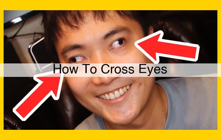
To cross eyes, focus on a point close to your nose and allow your eyes to inward. Gently relax your eyes and let them cross, as if looking at something very close. Gradually bring the object of focus closer until your eyes are fully crossed. Maintain focus on the object and avoid overexerting your eyes. If you experience any discomfort, stop the exercise and consult a medical professional.
Eye Movement Basics
The human eye is a remarkable organ, capable of complex and precise movements that allow us to navigate the visual world. These movements are controlled by six extraocular muscles, which work in concert to rotate and move the eyes in different directions.
- Superior rectus: Elevates the eye upward and inward.
- Inferior rectus: Depresses the eye downward and inward.
- Medial rectus: Adducts the eye (turns it inward) toward the nose.
- Lateral rectus: Abducts the eye (turns it outward) away from the nose.
- Superior oblique: Rotates the eye downward and outward.
- Inferior oblique: Rotates the eye upward and outward.
These muscles are innervated by various cranial nerves, including the trochlear (CN IV), abducens (CN VI), and oculomotor (CN III) nerves. The oculomotor nerve controls the superior rectus, medial rectus, inferior oblique, and levator palpebrae superioris muscles, which is responsible for opening the eyelids. The trochlear nerve innervates the superior oblique muscle, while the abducens nerve supplies the lateral rectus muscle.
The precise coordination of these muscles allows for smooth and controlled eye movements, enabling us to scan our surroundings, focus on objects, and maintain stability during head and body movements.
Binocular Vision
- Explain the processes of convergence and accommodation.
Binocular Vision: The Art of Precise Coordination
Binocular vision is the ability to use both eyes together to create a single, three-dimensional image. This complex process involves two key mechanisms: convergence and accommodation.
Convergence:
Imagine you’re looking at an object close to your face. To focus on the object, your extraocular muscles contract, causing both eyes to turn inward. This is convergence. The closer the object, the greater the convergence required.
Accommodation:
In addition to convergence, your lens also adjusts its shape to focus light on the retina. This process is called accommodation. When you look at something close, the lens bulges, making it thicker. When you look at something far away, the lens flattens.
The Interplay of Convergence and Accommodation
Convergence and accommodation work hand-in-hand. When you look at a near object, your eyes converge and your lens bulges. When you shift your gaze to a distant object, your eyes diverge (turn outward) and your lens flattens. This synchronized movement ensures that both eyes are focused on the same object at all times.
Benefits of Binocular Vision
Binocular vision provides numerous benefits, including:
- Depth perception: It allows us to perceive the distance and position of objects in three dimensions.
- Visual field: It provides a wider field of view than using one eye alone.
- Reduced eye strain: Proper binocular vision helps prevent eye fatigue and headaches.
- Binocular summation: Two eyes working together can detect objects more quickly and efficiently.
When Binocular Vision Goes Awry
In some cases, binocular vision can be impaired due to conditions such as strabismus, amblyopia, or diplopia. Strabismus occurs when the eyes are misaligned, resulting in crossed eyes or lazy eye. Amblyopia, also known as lazy eye, occurs when one eye is weaker than the other. Diplopia, or double vision, is a condition where the brain receives two different images from each eye, resulting in the perception of two overlapping objects.
Vision Impairments
- Define and discuss strabismus, amblyopia, and diplopia.
Vision Impairments: Common Eye Conditions and Their Impact
When our eyes don’t work quite as they should, it can significantly affect our vision and overall well-being. Here are three common vision impairments that you need to know about:
Strabismus (Crossed Eyes):
Imagine your eyes looking in different directions, creating a misalignment. That’s strabismus, commonly known as crossed eyes. It occurs when the muscles controlling eye movement are imbalanced, leading to a deviation in one or both eyes. Strabismus can be inward (esotropia), outward (exotropia), or even vertical. Persistent strabismus can cause double vision (diplopia) or even lazy eye (amblyopia).
Amblyopia (Lazy Eye):
When one eye is weaker than the other, amblyopia happens. The weaker eye fails to develop properly, often due to strabismus or other vision problems. Left untreated, amblyopia can lead to permanent vision loss in the affected eye. Usually, the brain favors the stronger eye, suppressing signals from the weaker one. Early detection and treatment are crucial to prevent vision loss.
Diplopia (Double Vision):
Seeing objects in duplicate is a frustrating experience! Diplopia, aka double vision, arises from a disruption in the coordination of the eye muscles. The eyes send different images to the brain, leading to a perceived doubling of objects. Diplopia can stem from nerve damage, eye muscle weakness, or even certain medical conditions such as stroke. Persistent double vision requires prompt medical attention to determine the underlying cause.
Nystagmus
- Describe the causes and symptoms of nystagmus.
Nystagmus: The Uncontrollable Eye Movement
Nystagmus, a condition characterized by involuntary and rhythmic back-and-forth movements of the eyes, can be a puzzling and disconcerting experience. These movements can occur horizontally, vertically, or in a circular pattern.
The causes of nystagmus are varied and can range from developmental factors to underlying medical conditions. Infantile nystagmus, the most common type, typically develops within the first six months of life and is associated with visual problems such as astigmatism or farsightedness. Other causes include acquired nystagmus, which may be triggered by neurological disorders like multiple sclerosis or traumatic brain injury, and sensory nystagmus, which arises from impaired vision in one eye.
The symptoms of nystagmus can be subtle or pronounced, depending on the severity of the condition. The most noticeable symptom is involuntary rapid eye movements. These movements can be constant or intermittent and can interfere with daily activities such as reading, writing, and driving. Other symptoms may include head nodding or tilting, which can be an attempt to stabilize vision, and poor depth perception.
If you experience nystagmus, it’s important to see an ophthalmologist or neurologist for evaluation. They will perform a comprehensive eye exam, including tests to assess your vision, eye movements, and neurological function. Based on the findings, they will determine the underlying cause and recommend appropriate treatment options.
In some cases, nystagmus can improve with simple measures such as wearing glasses or contact lenses to correct underlying vision problems. In other cases, more specialized treatments may be needed, such as drug therapy to control eye movements or eye muscle surgery to improve eye alignment.
Living with Nystagmus
While nystagmus can present challenges, it’s important to remember that it does not affect intelligence or overall life expectancy. With proper diagnosis and management, individuals with nystagmus can lead full and productive lives.
Support and Resources for Individuals with Nystagmus
Several organizations provide support and resources for individuals with nystagmus and their families. These organizations offer information on the condition, coping strategies, and access to medical professionals specializing in nystagmus treatment.
By understanding the causes, symptoms, and treatment options for nystagmus, you can empower yourself to manage this condition and live a comfortable and fulfilling life.
Nerve Damage and Its Impact on Eye Movement
Understanding the Role of Nerves in Eye Movement
Nerves play a crucial role in controlling the movement of our eyes, allowing us to focus on objects, track moving targets, and maintain stable vision. These cranial nerves originate in the brain and connect to muscles responsible for eye movement.
Potential Causes of Nerve Damage
1. Trauma: A blow or injury to the head or eye socket can damage nerves responsible for eye movement, leading to double vision, gaze palsy, and other vision problems.
2. Infections: Infections such as meningitis or syphilis can cause inflammation and damage to nerves, affecting eye movement.
3. Neurological Diseases: Conditions like multiple sclerosis and Parkinson’s disease disrupt nerve function, which can manifest in eye movement problems.
4. Diabetes: Over time, diabetic neuropathy can damage the nerves controlling eye muscles, leading to vision issues and possible eye muscle weakness.
Consequences of Nerve Damage on Eye Movement
Damage to eye nerves can result in various eye movement disorders, including:
- Strabismus (Crossed Eyes): One eye may turn inward or outward, causing double vision.
- Gaze Palsy: The eyes lose the ability to move in certain directions, limiting vision and causing difficulty in reading and driving.
- Nystagmus: Involuntary, rhythmic eye movements, often caused by brain or nerve damage.
- Ptosis (Droopy Eyelid): Nerve damage to the muscle that lifts the eyelid can cause the eyelid to droop, obstructing vision.
Importance of Seeking Medical Attention
If you notice any changes in your vision or eye movement, it’s crucial to seek medical attention promptly. Early diagnosis and treatment can prevent permanent vision problems and improve overall eye health. Your healthcare provider will perform comprehensive eye exams and may recommend further tests, such as MRI or CT scans, to identify the underlying cause of nerve damage and determine appropriate treatment options.
Muscular Weakness and Its Impact on Eye Movement
Eye movements are essential for everyday tasks such as reading, driving, and navigating our environment. These movements are controlled by a complex system of muscles that work together seamlessly. However, muscular weakness in these muscles can significantly impair eye movement, leading to various vision problems.
Causes of Muscular Weakness:
Muscular weakness in the eye can stem from a variety of causes, including:
- Myasthenia gravis: An autoimmune disorder that weakens muscles throughout the body, including the eye muscles.
- Thyroid eye disease: A condition that affects the immune system and causes inflammation in the muscles around the eyes.
- Neurological disorders: Certain neurological conditions, such as Parkinson’s disease and multiple sclerosis, can affect the nerves that control eye movement.
- Muscular dystrophies: A group of genetic disorders that cause progressive muscle weakness and wasting.
- Trauma: Eye muscle injuries, such as those caused by surgery or blunt force, can also lead to weakness.
Effects on Eye Movement:
Muscular weakness can affect eye movement in several ways:
- Ptosis: Drooping of the upper eyelid due to weakness of the levator palpebrae muscle.
- Strabismus: Misalignment of the eyes, where one eye turns inward, outward, upward, or downward due to weak muscles that control the eye’s position.
- Nystagmus: Involuntary, rapid eye movements caused by weakness in the muscles that stabilize eye position.
- Reduced eye motility: Limited range of motion of the eyes due to weak muscles responsible for moving the eyes in different directions.
Diagnosis and Treatment:
Proper diagnosis of muscular weakness in the eye is crucial for determining the underlying cause and providing appropriate treatment. Eye exams, imaging tests, and nerve function tests may be used to identify the exact condition. Treatment options may include:
- Medications: To manage symptoms and strengthen muscles in certain conditions, such as myasthenia gravis.
- Prisms: Lenses that are placed in front of the eyes to help correct strabismus.
- Eye muscle surgery: A surgical procedure to reposition or strengthen weakened eye muscles.
- Physical therapy: To improve muscle coordination and strengthen the muscles surrounding the eyes.
Diagnostic Tests
- Describe different tests used to assess eye movement and visual function.
Diagnostic Tests for Eye Movement and Visual Function
Navigating the intricate world of eye movement and visual function requires a thorough evaluation. Diagnostic tests provide invaluable insights into potential impairments, paving the way for accurate diagnoses and appropriate treatment plans.
Ocular Motility Examination:
This comprehensive assessment evaluates the range and precision of eye movements. The examiner observes horizontal, vertical, and diagonal movements, as well as the ability to follow a moving target. Abnormal eye movements, such as nystagmus (involuntary eye oscillations), can indicate underlying neurological or muscular issues.
Cover Test:
This simple yet effective test screens for misalignment of the eyes, known as strabismus. The examiner covers one eye while the patient looks straight ahead, revealing any deviation in the uncovered eye’s position. Squinting or a head turn may be compensatory mechanisms to overcome misalignment.
Stereopsis Test:
Stereopsis is the ability to perceive depth. Using special glasses or tests, this assessment evaluates the patient’s three-dimensional vision, which is crucial for everyday activities like driving and sports. Impaired stereopsis can result from eye muscle coordination issues or certain eye conditions.
Visual Acuity Test:
This familiar test measures the eye’s ability to focus on objects at various distances. It involves reading letters or symbols on a chart, and the results are expressed as a fraction (e.g., 20/20). Reduced visual acuity can indicate refractive errors (e.g., nearsightedness or farsightedness), cataracts, or other eye disorders.
Visual Field Test:
This test assesses the patient’s peripheral vision, or the range of what they can see to the sides while looking straight ahead. It helps detect blind spots or scotomas, which may be caused by damage to the retina, optic nerve, or certain neurological conditions.
Electrophysiological Tests:
These specialized tests use electrodes to record the electrical activity of the retina (electroretinography) and optic nerve (electro-oculography). They can identify abnormalities in nerve function, which may be associated with conditions like glaucoma, optic neuritis, or multiple sclerosis.
MRI and CT Scans:
Advanced imaging techniques like magnetic resonance imaging (MRI) and computed tomography (CT) scans provide detailed images of the eyes and surrounding structures. They can reveal tumors, bleeding, or other abnormalities that may affect eye movement or visual function.
By employing these diverse diagnostic tests, ophthalmologists can gain a comprehensive understanding of a patient’s eye movement and visual function. These tests empower healthcare professionals to make informed diagnoses and tailor customized treatment plans to restore optimal eye health.
Eye Muscle Surgery: Restoring Ocular Alignment
When eye muscles malfunction, causing misalignment in the eyes, corrective surgery may be necessary to restore proper vision. Eye muscle surgery, also known as strabismus surgery, aims to adjust the position and tension of the six extraocular muscles that control eye movement.
Indications for Eye Muscle Surgery:
- Strabismus: Crossed eyes (esotropia) or outward-turned eyes (exotropia) can be corrected through surgery to align the eyes properly.
- Diplopia: Double vision caused by misaligned eyes can be alleviated by surgical adjustments to the eye muscles.
- Vertical misalignment: Surgery can correct vertical eye misalignment, such as when one eye is higher or lower than the other, resulting in difficulties with depth perception and reading.
Surgical Approach:
Eye muscle surgery is typically performed under general anesthesia. The surgeon makes a small incision in the conjunctiva, the clear membrane covering the white of the eye, to access the extraocular muscles. Using fine sutures, the surgeon adjusts the muscle’s insertion point on the eye, altering its tension and position.
The goal of surgery is to create a balanced muscle system that allows the eyes to move together in a coordinated manner. The exact surgical technique used depends on the type and severity of the eye misalignment.
Recovery from Eye Muscle Surgery:
After surgery, the patient’s eyes will be patched for a few days to promote healing. Mild discomfort and swelling are possible, which can be managed with pain relievers. Most patients notice an immediate improvement in eye alignment, but complete recovery may take several weeks.
Benefits of Eye Muscle Surgery:
- Improved vision: Surgery can correct double vision and misalignment, resulting in enhanced visual clarity.
- Enhanced depth perception: Proper eye alignment enables better depth perception for activities like driving and sports.
- Reduced fatigue: Misaligned eyes can cause eye strain and fatigue, which surgery can alleviate.
- Improved facial aesthetics: Eye muscle surgery can address misaligned eyes that may be cosmetically unappealing, restoring a more balanced appearance.
Eye muscle surgery is a highly effective and safe procedure to correct eye misalignment. By adjusting the position and tension of the extraocular muscles, surgery improves visual function, depth perception, and overall eye health.


