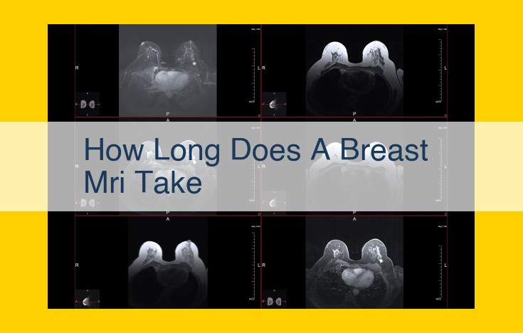
The duration of a breast MRI varies based on multiple factors, including patient age, health history, breast size and density, type of sequence used, contrast agent administration, MRI scanner strength, and scheduling efficiency. Patient preparation and comfort level, as well as the specific slice thickness, field of view, and sequences employed, can also impact scan time. Standardized protocols and efficient scheduling practices help optimize the process. Delays or interruptions can occasionally extend scan duration.
Age and Health History:
The age of the patient can influence MRI scan duration. Elderly patients tend to have longer scan times due to age-related changes in their physiology and body composition. Their tissues may have reduced water content, leading to longer signal acquisition times.
Health history also plays a role. Patients with certain medical conditions, such as cardiovascular disease or claustrophobia, may require additional preparation or modifications to the scan protocol, potentially extending the scan duration.
Patient Preparation and Comfort:
Adequate preparation can significantly reduce scan time. Patients should:
- Arrive on time to allow for necessary paperwork and preparation.
- Fast and hydrate as instructed to ensure clear images.
- Wear comfortable clothing that allows for easy positioning.
Patient comfort is crucial. A relaxed patient is more cooperative, leading to a smoother scan and potentially shorter scan times. Providing noise-canceling headphones, blankets, and pillows can enhance comfort.
Imaging-Related Factors Affecting MRI Scan Duration: A Detailed Exploration
Magnetic resonance imaging (MRI) scans are essential diagnostic tools that provide detailed images of the body’s internal structures. The duration of an MRI scan can vary depending on a host of factors, including those related to the imaging procedure itself. In this post, we dive into the imaging-related elements that influence scan time, helping you better understand the process and its implications.
1. Breast Size and Density
Breast size and density play a significant role in determining MRI scan duration. Larger breasts require more time to scan, as the scanner must cover a wider area. Dense breasts, which contain more glandular tissue, also require longer scan times to obtain clear images.
2. Type of Sequence
MRI scans utilize various sequences to produce different types of images. T1-weighted sequences, commonly used for evaluating anatomy, tend to have shorter scan times. T2-weighted sequences, which highlight fluid-filled structures, typically require longer scan times. Additionally, specialized sequences like diffusion-weighted imaging (DWI) can add to the overall scan duration.
3. Slice Thickness
The slice thickness refers to the width of each image slice captured during the MRI scan. Thinner slices provide more detailed images but require a longer scan time. Conversely, thicker slices cover a larger area in less time but may compromise image quality.
4. Field of View (FOV)
The field of view (FOV) determines the size of the anatomical area being scanned. A larger FOV, encompassing a wider region, requires more time to scan. Adjusting the FOV appropriately can optimize scan time without sacrificing image quality.
5. Contrast Agent
Contrast agents, injected into the bloodstream, enhance the visibility of certain tissues and lesions. The type, dosage, and timing of contrast agent injection can all impact scan duration. Some contrast agents require additional time for the substance to distribute throughout the body before imaging can commence.
How Different MRI Sequences Affect Scan Time
Magnetic Resonance Imaging (MRI) is a non-invasive imaging technique that uses powerful magnets and radio waves to create detailed pictures of the inside of the body. The duration of an MRI scan can vary depending on several factors, including the type of MRI sequence used.
T1-Weighted Imaging
T1-weighted imaging (T1WI) is a type of MRI sequence that creates images based on the relaxation time of protons in the body. Protons in different tissues relax at different rates, so T1WI can be used to differentiate between different types of tissue. T1WI scans are typically faster than other MRI sequences because they require fewer repetitions.
T2-Weighted Imaging
T2-weighted imaging (T2WI) is another type of MRI sequence that creates images based on the relaxation time of protons in the body. However, T2WI scans are more sensitive to water content than T1WI scans. This means that T2WI scans can provide better contrast between different types of soft tissue, but they take longer to acquire than T1WI scans.
Diffusion-Weighted Imaging
Diffusion-weighted imaging (DWI) is a type of MRI sequence that measures the diffusion of water molecules in the body. DWI scans can be used to detect abnormalities in the brain and other organs that may not be visible on other MRI sequences. DWI scans are typically longer than T1WI and T2WI scans because they require more repetitions.
Choosing the Right MRI Sequence
The type of MRI sequence that is used for a particular scan will depend on the clinical question being asked. For example, T1WI scans are often used to image the brain and spine, while T2WI scans are often used to image the腹部 and pelvis. DWI scans are often used to image the brain and other organs that may be affected by cancer or stroke.
By understanding the different types of MRI sequences and their varying scan times, patients can be better prepared for their MRI scans and can ask informed questions about the procedure.
Contrast Agent Considerations: Impact on MRI Scan Duration
When undergoing an MRI scan, the injection of a contrast agent plays a crucial role in optimizing the visibility and clarity of certain body tissues. However, the type, dosage, and timing of the contrast agent injection can also significantly influence the duration of the scan.
Type of Contrast Agent
MRI contrast agents are typically classified into two main categories: gadolinium-based contrast agents (GBCAs) and non-gadolinium-based contrast agents (NGCAs). GBCAs are the most commonly used, enhancing the visibility of blood vessels and certain organs, while NGCAs are often used as an alternative for patients with allergies or concerns about potential side effects from GBCAs. The choice of contrast agent can affect the scan time, with some agents requiring longer acquisition times due to their slower distribution throughout the body.
Dosage of Contrast Agent
The dosage of contrast agent administered is another factor that can influence scan duration. The higher the dosage, the more intense the signal enhancement will be in the targeted tissues. However, it’s important to note that higher dosages may also increase the time required for the contrast agent to clear from the body, potentially extending the scan duration.
Timing of Contrast Agent Injection
The timing of the contrast agent injection is a critical consideration. In general, the contrast agent is injected shortly before the acquisition of the targeted images, allowing it to reach the desired tissues and provide optimal enhancement. The scan duration can vary depending on the specific imaging protocol and the tissue being visualized. For example, scans involving the heart or abdominal organs may require multiple contrast agent injections and longer scan times to capture the desired images.
Additional Considerations
In addition to the factors mentioned above, certain conditions and patient characteristics can also affect the duration of the MRI scan with contrast. These may include:
- Patient’s body weight and size
- Allergies or sensitivities to contrast agents
- Underlying health conditions or kidney function
- Patient’s tolerance and comfort level
By carefully considering the type, dosage, and timing of contrast agent injection, radiologists can optimize the MRI scan duration while ensuring the highest quality of images for accurate diagnosis and treatment planning.
Technical Equipment Factors and Their Impact on MRI Scan Duration
When it comes to MRI scans, the precision of the images and the speed at which they can be acquired depend heavily on the technical equipment used. Here’s how three key factors – MRI scanner strength, coil type, and software – play a crucial role in determining MRI scan duration:
MRI Scanner Strength:
MRI scanners are characterized by their magnetic field strength, measured in Tesla (T). The strength of the magnetic field directly influences the signal-to-noise ratio (SNR) of the images. A stronger magnetic field generally leads to a higher SNR, resulting in clearer images. However, higher field strengths also require longer scan times to achieve optimal image quality.
Coil Type:
MRI coils are responsible for transmitting and receiving radiofrequency signals from the patient’s body. Different types of coils are designed for specific body regions and can significantly impact scan time. For example, surface coils, which are placed directly on the skin, provide excellent SNR but have a smaller field of view, resulting in shorter scan times. On the other hand, volume coils, which cover a larger area, offer a wider field of view but may require longer scan times to achieve the same SNR.
Software:
MRI software plays a critical role in optimizing scan parameters and reconstruction algorithms. Advanced software techniques can reduce scan time by utilizing parallel imaging techniques, which allow for simultaneous acquisition of data from multiple coils. Additionally, software can enhance image quality by reducing noise and artifacts, which may lead to shorter scan times or the ability to acquire higher-quality images in the same amount of time.
By understanding the influence of these technical equipment factors, radiologists and MRI technologists can tailor scan protocols to achieve the optimal balance between image quality and scan duration, ensuring efficient and effective MRI examinations.
Standardization and Protocols: The Key to Efficient MRI Scans
When it comes to MRI scans, standardized protocols play a crucial role in ensuring optimal scan duration. By establishing clear guidelines for each step of the MRI process, medical professionals can eliminate unnecessary delays and ensure that scans are completed as efficiently as possible.
For instance, standardizing patient preparation can save significant time. Clear instructions on how to dress, remove metal objects, and lie still during the scan enable patients to cooperate fully, reducing the need for rescans due to motion artifacts.
Moreover, protocol standardization for imaging parameters is essential. By defining the optimal sequence types, slice thicknesses, and field of view for different examinations, technicians can avoid having to adjust settings repeatedly, saving valuable time.
Furthermore, the use of pre-defined protocols for contrast agent administration ensures that patients receive the correct dosage and timing, eliminating delays caused by unnecessary injections or repeated scans due to inadequate contrast enhancement.
In conclusion, adhering to standardized protocols throughout the MRI process not only improves scan efficiency but also enhances the overall quality of the images. By streamlining the procedure and minimizing potential delays, technicians and radiologists can focus on providing high-quality diagnostic information while ensuring that patients experience a comfortable and efficient scan process.
Scheduling-Related Factors That Impact MRI Scan Duration
The Importance of Efficient Scheduling
The duration of an MRI scan can be significantly influenced by scheduling-related factors, including:
-
Number of patients scheduled: Overbooking can lead to delays as technicians struggle to keep up with the demand. Optimize scheduling to ensure a manageable patient load and minimize waiting times.
-
Availability of staff: Ensure adequate staffing levels to avoid technician shortages that can cause delays. Skilled technicians are essential for efficient scan execution and accurate image interpretation.
-
Scheduling efficiency: Implement effective scheduling practices that minimize downtime and optimize equipment utilization. Consider staggering appointments and using appointment reminders to reduce patient no-shows or late arrivals.
By addressing these scheduling factors, healthcare facilities can streamline the MRI scanning process, reduce patient wait times, and improve overall efficiency.
Other Factors that Impact MRI Scan Duration
Beyond the well-known factors that influence MRI scan time, several unforeseen circumstances can also affect the duration of the procedure. These include:
Technical Delays
MRI scanners, like any other complex machinery, can occasionally experience technical glitches or malfunctions. These delays can disrupt the scanning process, extending the overall time required to complete the exam.
Interruptions
Unforeseen interruptions, such as the need for a radiologist’s consultation or an urgent patient call, can also lead to delays in the MRI scan. These interruptions can break the scanning flow and require additional time for setup and preparation.
Patient Positioning
In certain cases, patients may experience difficulty maintaining proper positioning throughout the MRI scan. This can result in the need for repositioning or additional scans, which can prolong the examination time.
Movement
Even involuntary patient movement, such as breathing or swallowing, can affect the quality of MRI images. In these situations, the scan may need to be repeated or additional time allocated to compensate for motion artifacts.
Delayed Contrast Enhancement
For MRI exams that involve the use of a contrast agent, the timing of the injection can impact scan duration. If the patient experiences delayed contrast enhancement, which can be caused by factors such as patient weight or hydration status, the scan may need to be extended to allow for optimal visualization of the targeted tissues.
Understanding these additional factors can help patients and healthcare professionals better anticipate and manage the potential variability in MRI scan times. By optimizing the scanning process and minimizing unforeseen delays, we can ensure efficient and accurate MRI examinations that provide the best possible diagnostic information.
